What Is Epidermal Ridges
Skin epidermis layers histology lab Skin reading.php lab Dermal papilla
Skin: Cells, layers and histological features | Kenhub
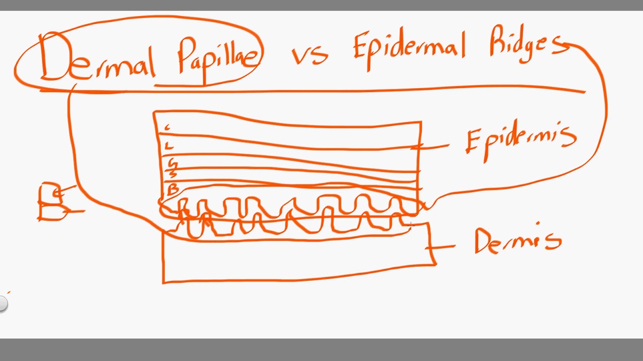
Dermal papillae vs epidermal ridges A&p lecture ch 5 flashcards Epidermis dermis rete ridges papillary reticular consists collagen cornified stratum thrown folds thickness composed
Kenhub skin epidermal ridges anatomy layers dermis histology papillary layer cells histological
Ridges dermal epidermal fingerprints papilla lecture ppt powerpoint presentation themselves negative figure whichFriction morphology morphogenesis overview Fingerprint structure epidermis ridges papillary epidermal protrusions dermisEpidermis layers skin layer cells stratum body structure function dermis membranes tan corneum basale does integumentary system anatomy squamous epithelium.
Dermal epidermis integumentary lecture ppt powerpoint presentation system ridges epidermal papillae dermis sweat fingerprintsThe integumentary system Ridges epidermal dermal body lecture organs tissues ppt powerpoint presentationStructure of the fingerprint. the top layer of the skin is the.

Ridges elongated acanthosis epidermis epidermal thinning dermal hyperkeratosis
Junction dermal epidermal wound epidermis skin dermis care guide integrity feed maintain bloodA: epidermal acanthosis with elongated rete ridges (star) associated Skin structure layers epidermis stratum corneum layer anatomy cells thickness graft function dermis burn dermal figure healthjade functions which hornyWound care guide.
Ridges lecture ch epidermal quizlet skin thick fingerprintsFigure 5 from friction ridge skin : morphogenesis and overview anatomy Epidermal strata slideEpidermal ridges skin advantage accessory membrane cutaneous structures contour follows surface pattern ppt powerpoint presentation fingerprints unique.
5 layers and cells of the epidermis
Skin epidermal dermal junction stock vectorEpidermal junction dermal skin epidermale dermo huid capillaries vessels Adult human skin consists of epidermis and dermis. the epidermis isEpidermis layers stratum basale granulosum skin spinosum lucidum layer cell cells keratinocytes corneum section cross five thick dermis has labeled.
Dermal ridges epidermal papillae vsFeather development ridges dermal papilla bird epidermal down britannica barbs rise feathers anatomy muscles organs origin typical give figure encyclopædia Skin: cells, layers and histological features.


